for permission information see the print copy of You Can’t Just Snap Out of It
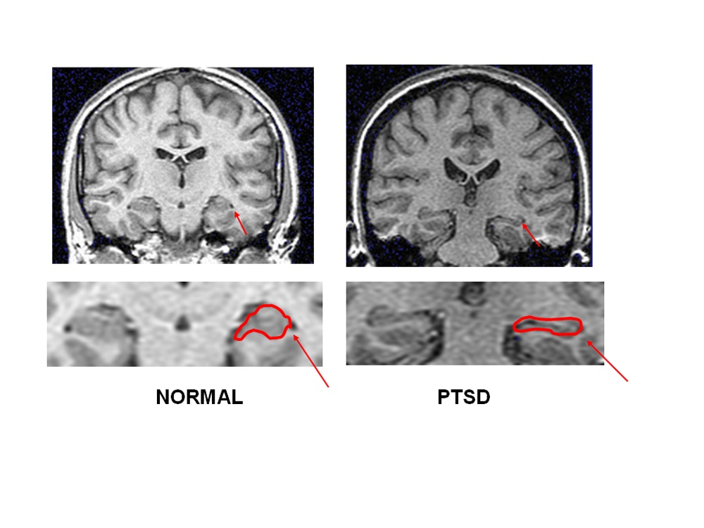
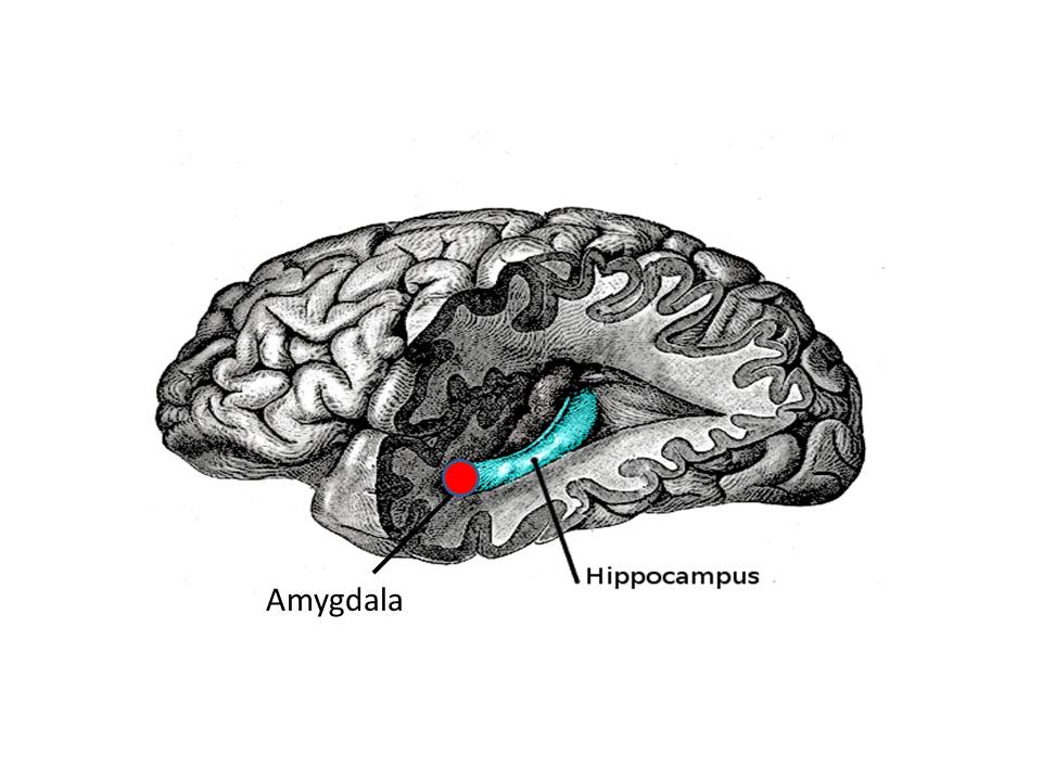
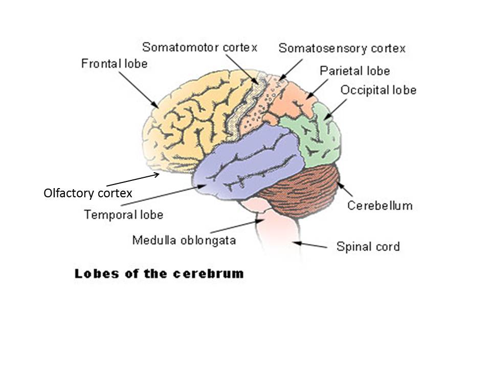
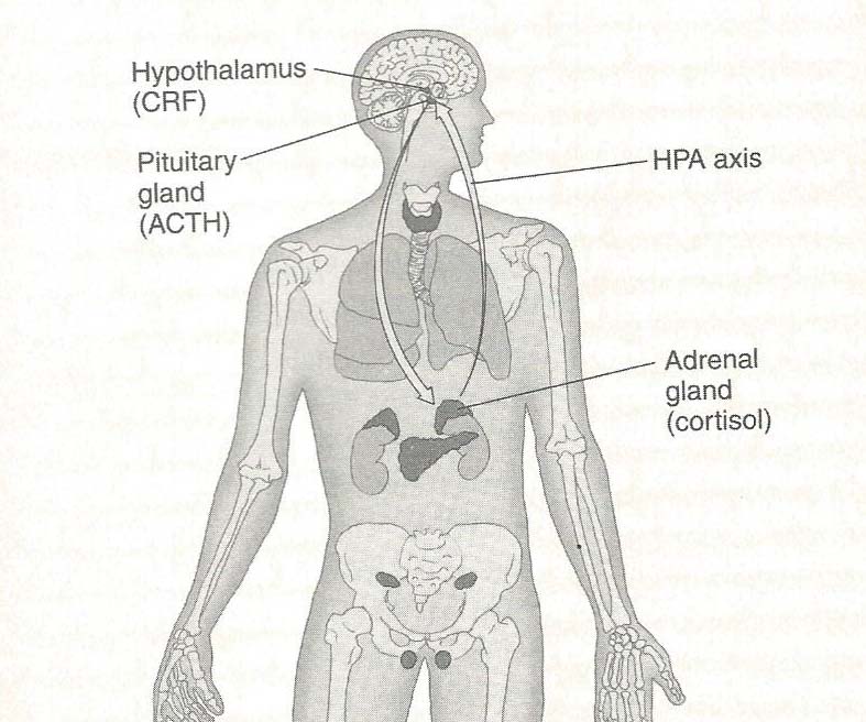
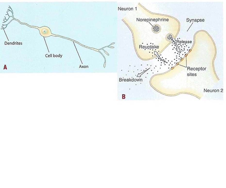
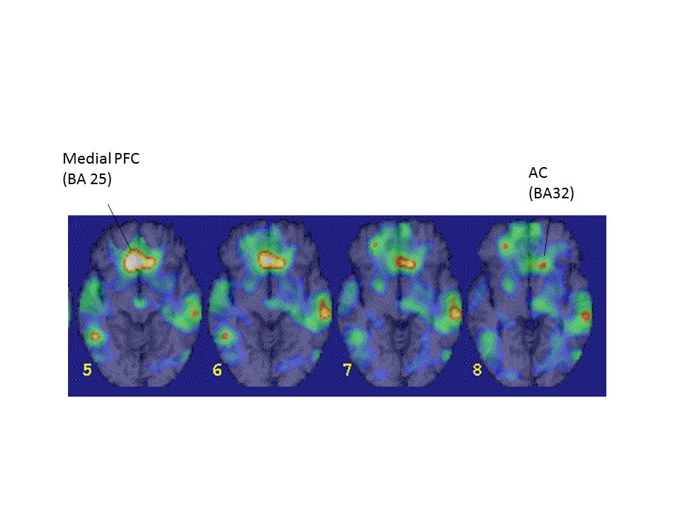
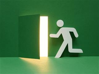
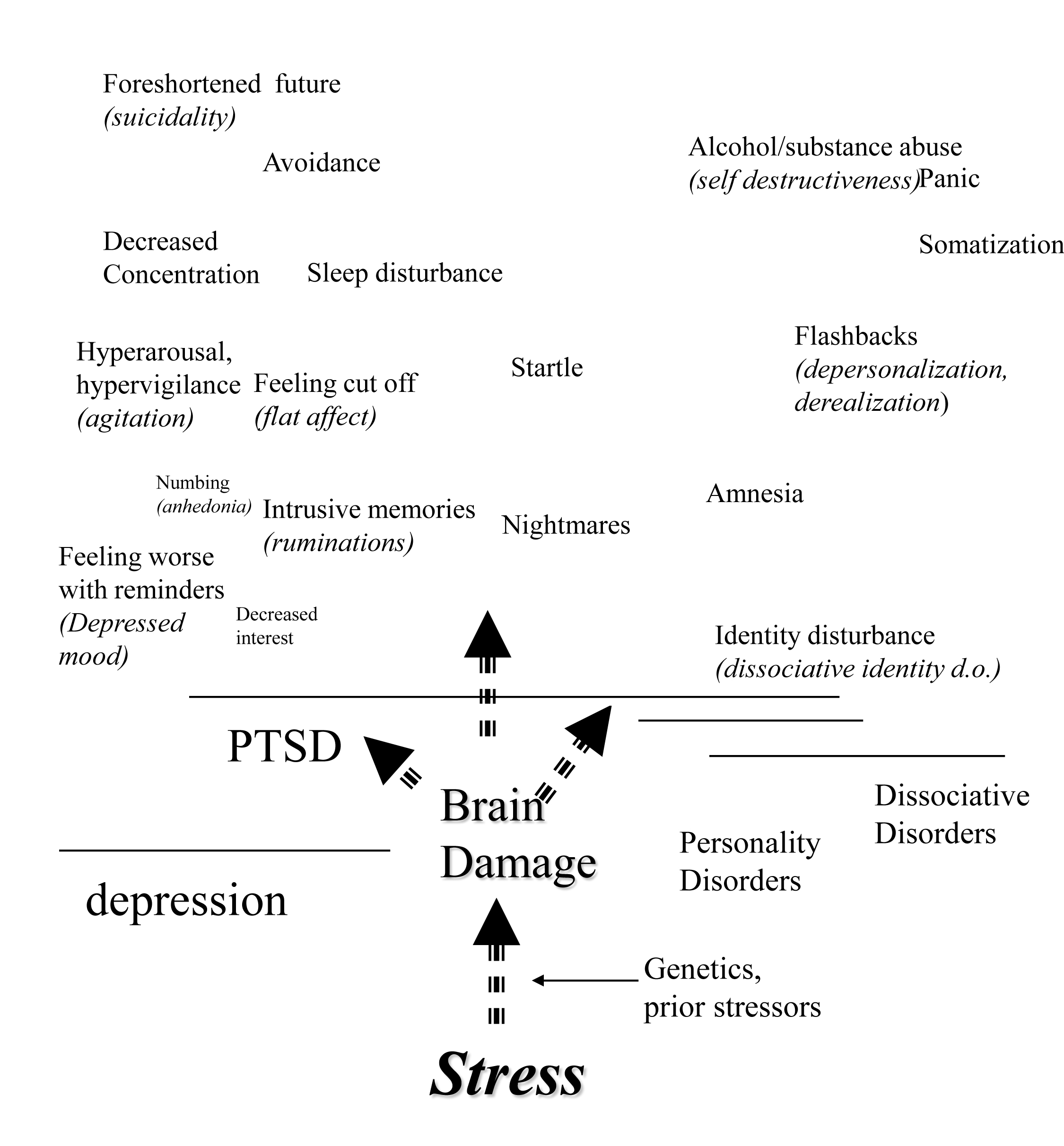
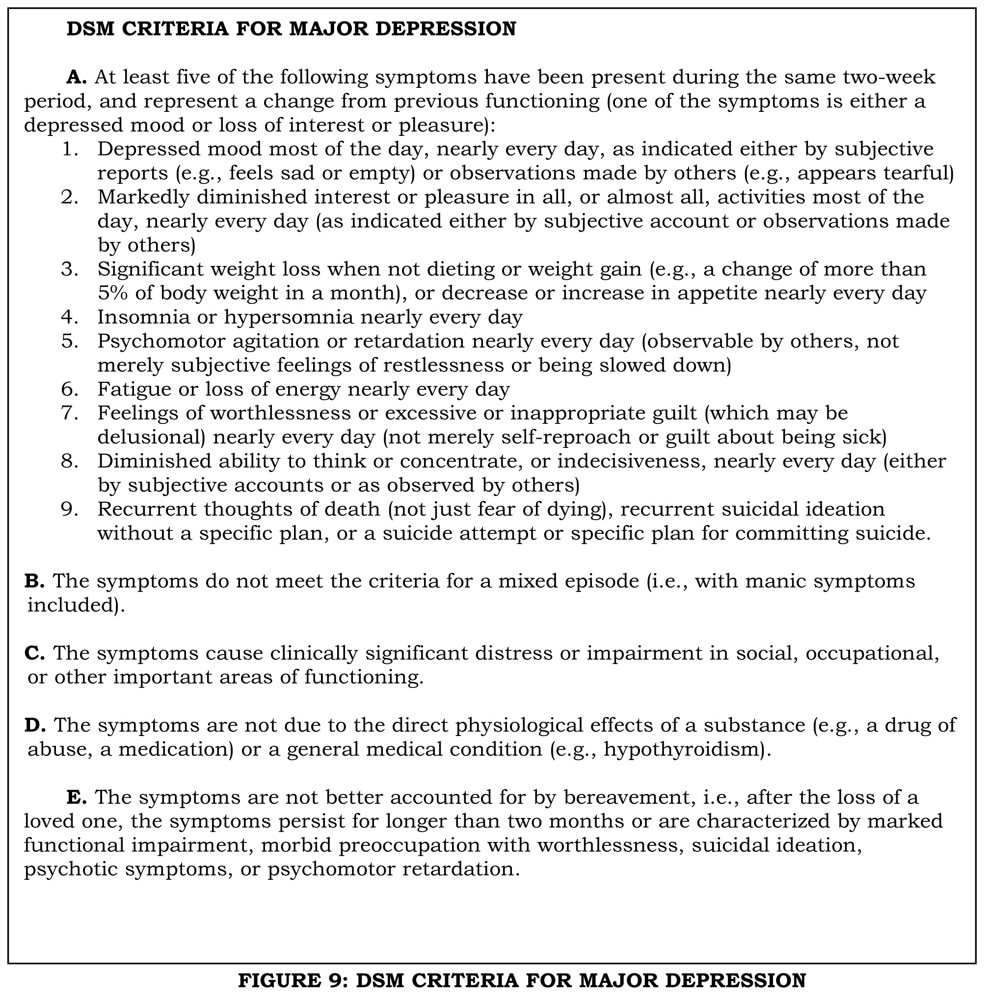
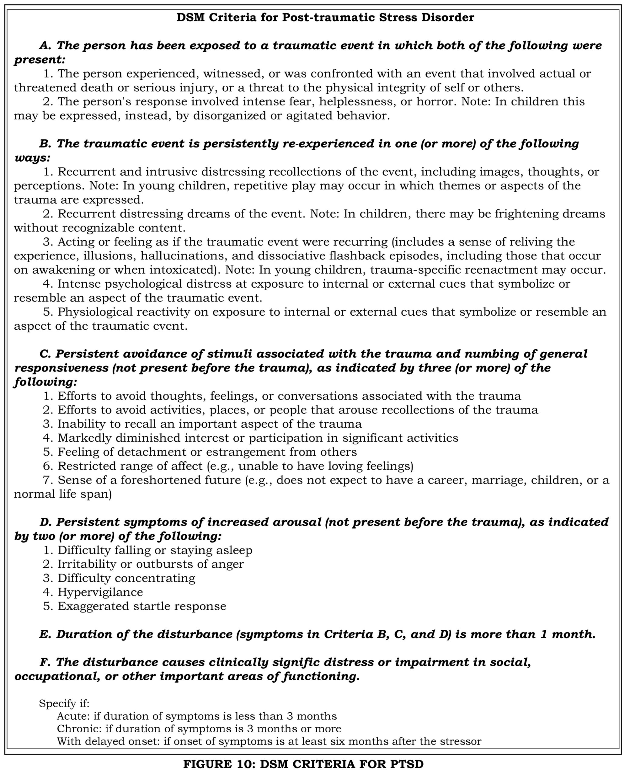
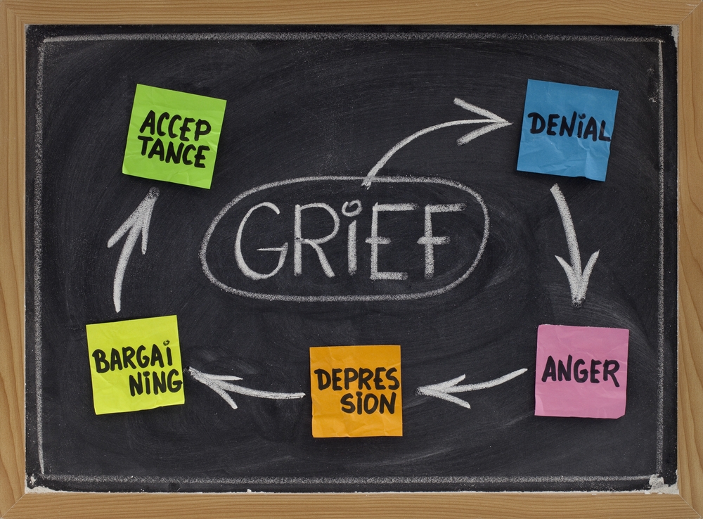
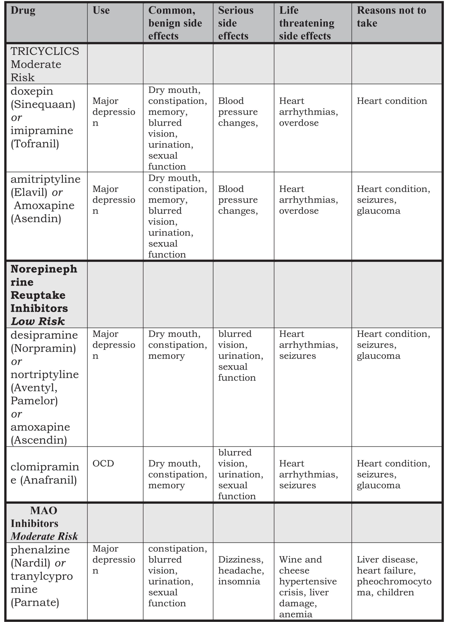
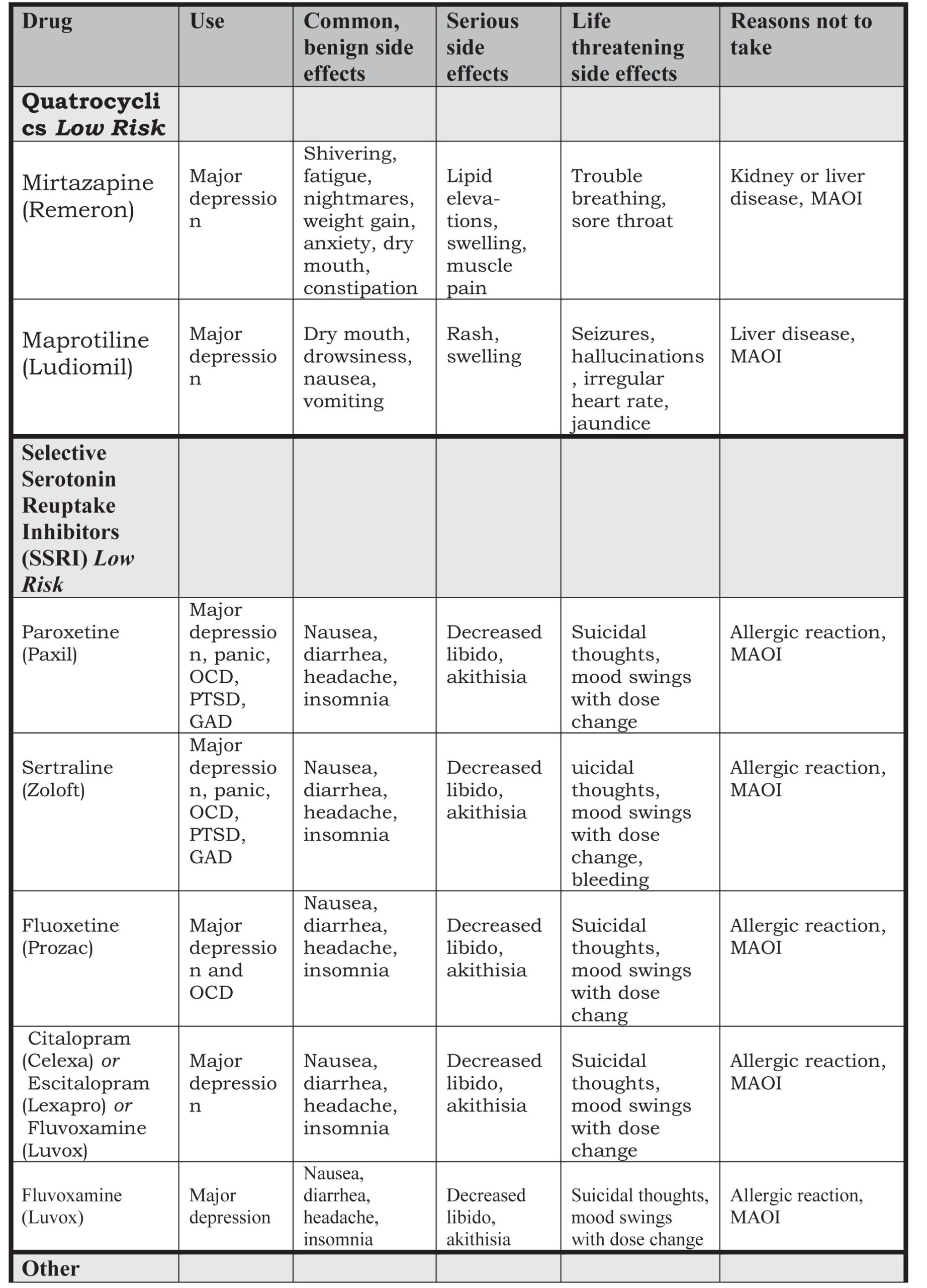
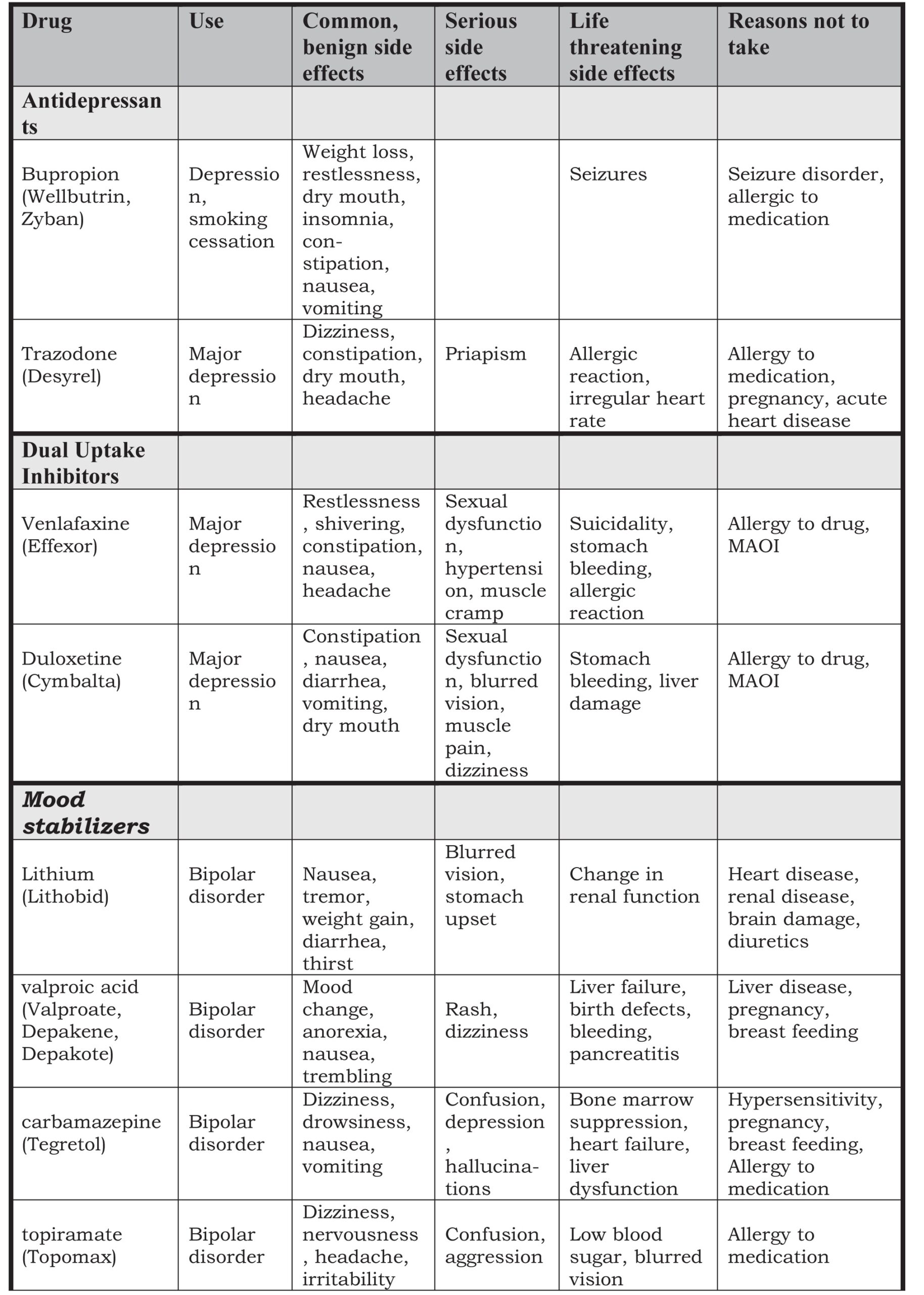
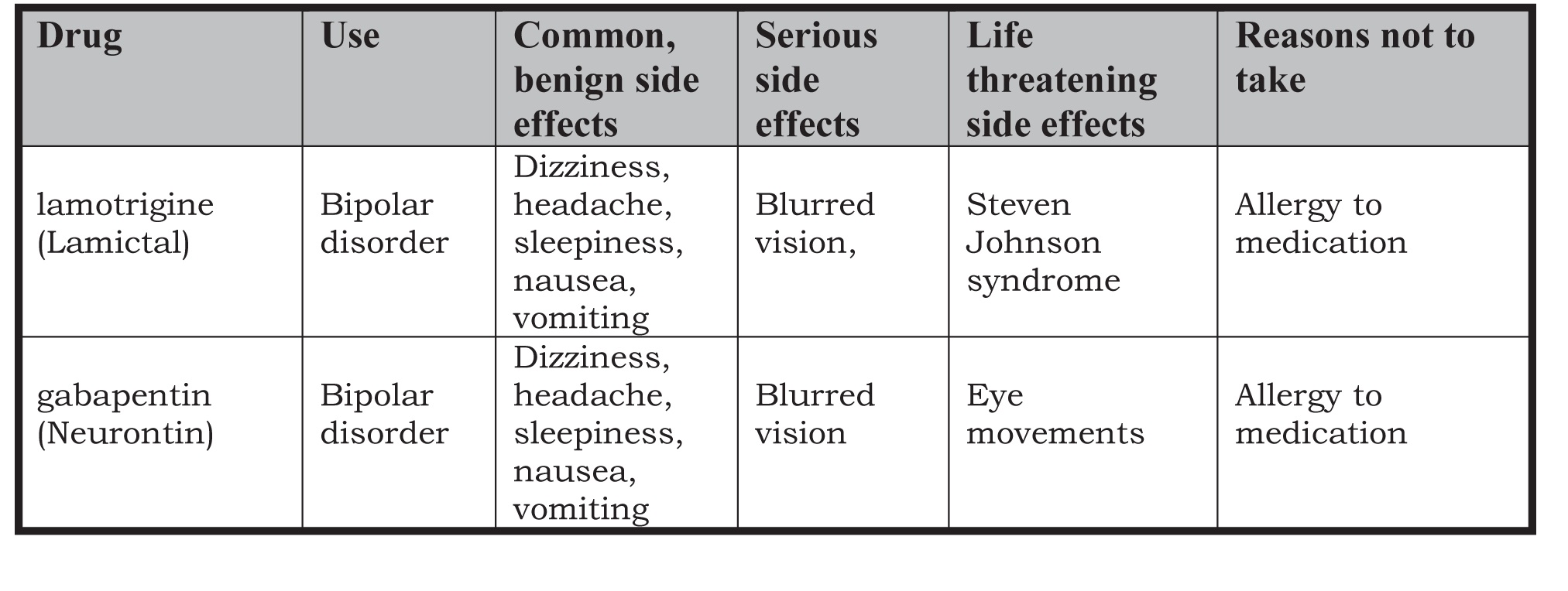
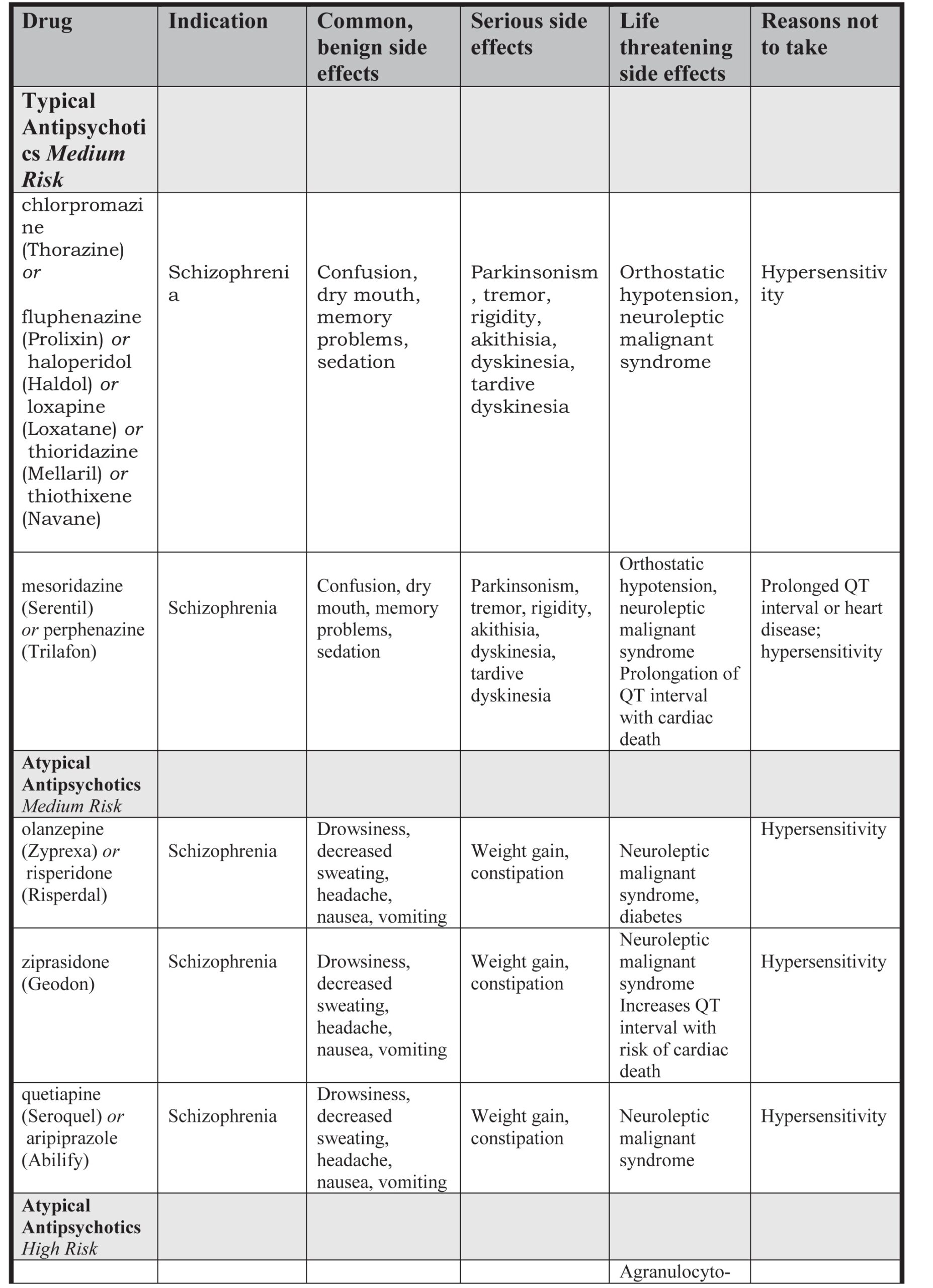
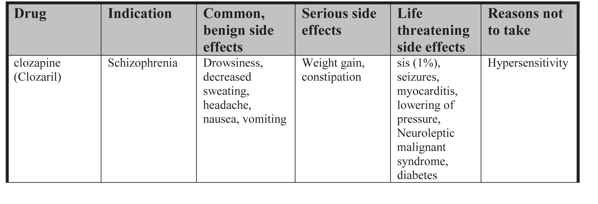
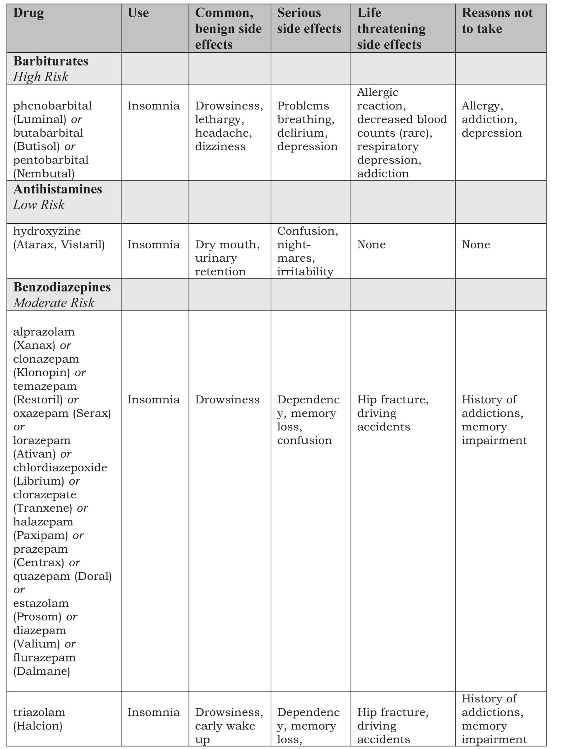
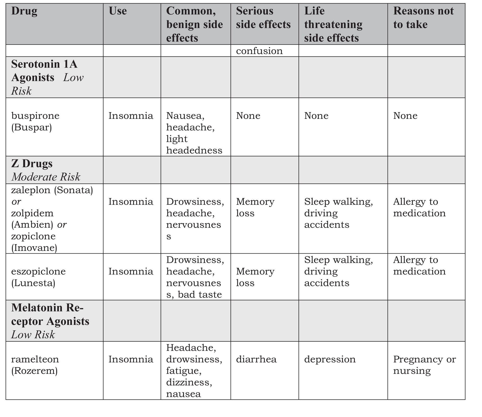
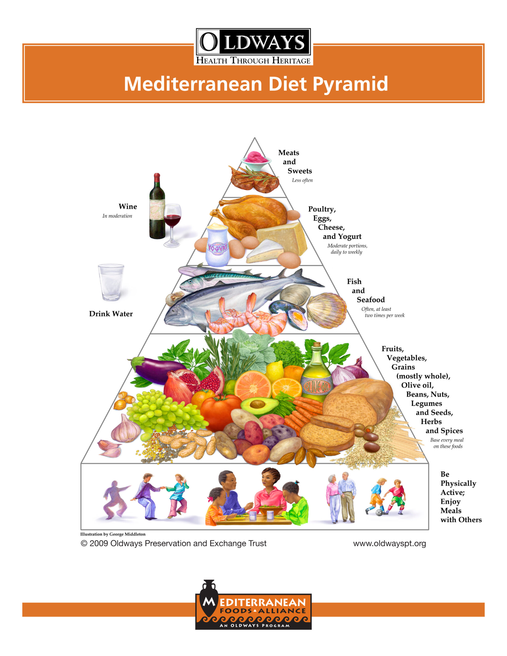
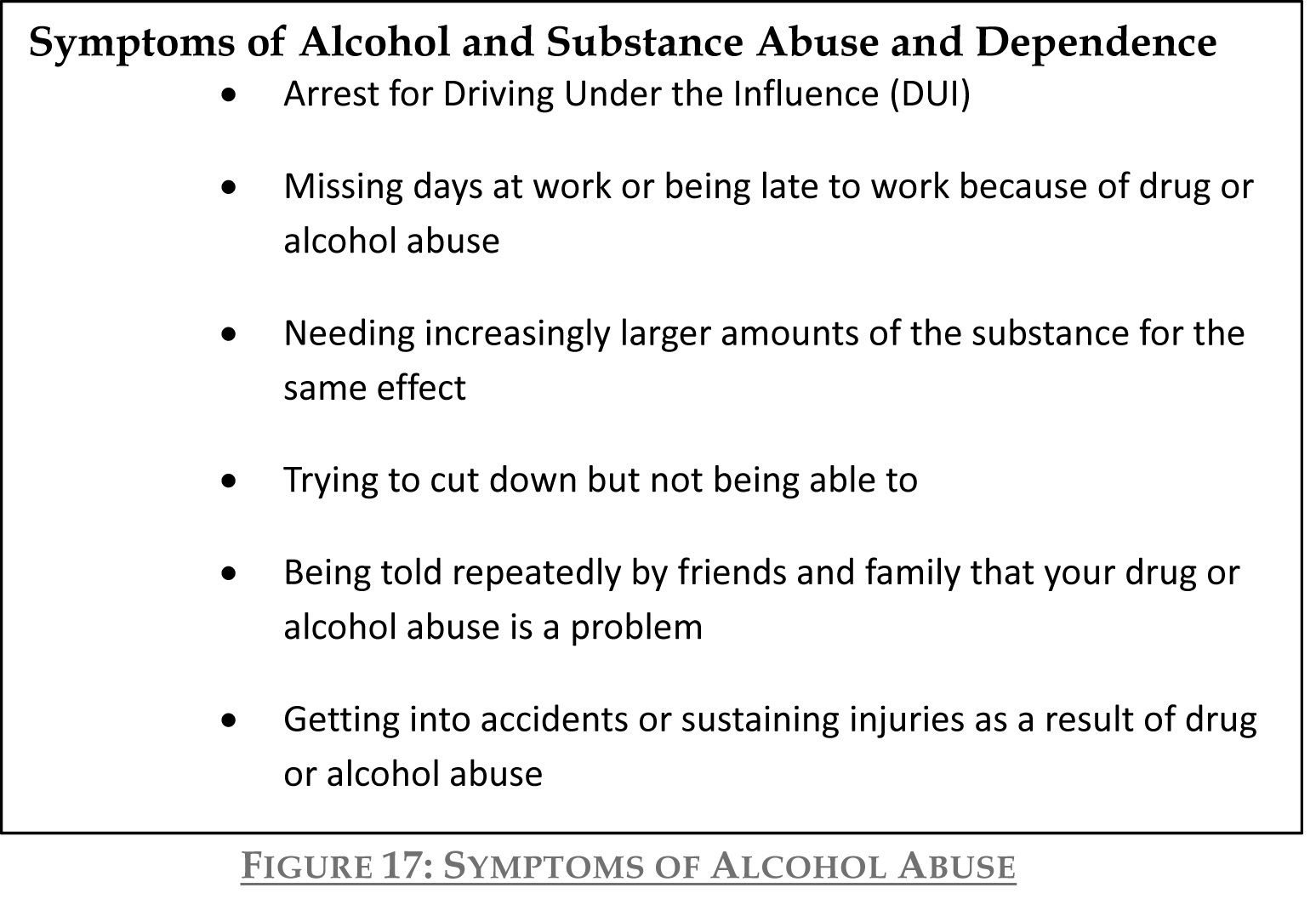
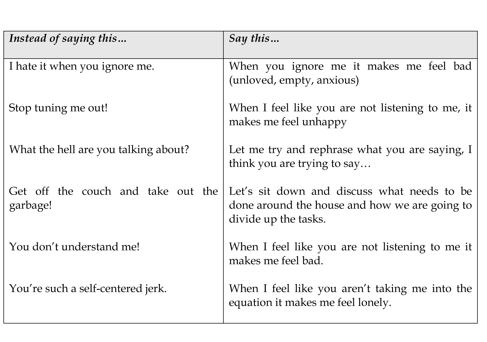
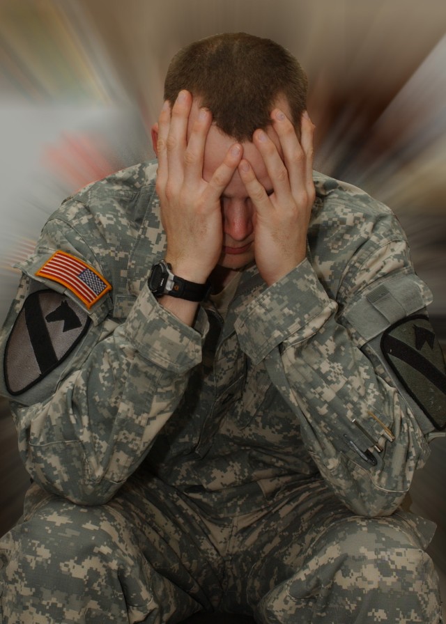
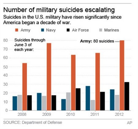
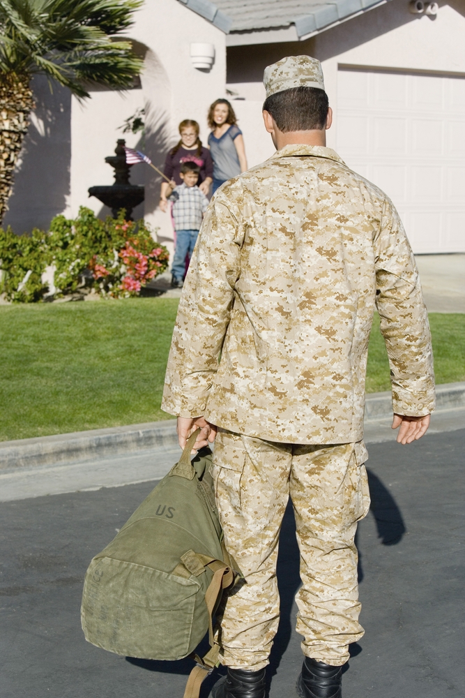
Figure 1. Hippocampal volume measured with magnetic resonance imaging (MRI). There is a visible reduction in the hippocampus (outlined in red) in a patient with PTSD compared to non-PTSD (reused with permission). Figure 2. Diagram of the human brain showing the amygdala (red) and hippocampus (blue). The frontal lobe (front part of the brain) is to the left of the diagram. The hippocampus and amygdala are on both the left and right sides of the brain (Source: Wikipedia). Figure 3. The cerebrum, or cerebral cortex, is at the outer part of the Figure 4. Neurohormonal Responses to Stress. Each of the body glands produces and releases Figure 5 . Diagram of a neuron, the basic cell of the brain. Long processes called axons release neurotransmitters into the space between neurons, called the synapse, where they travel to the next neurons. SSRIs work by blocking the reuptake of serotonin thus effectively increasing the amount of neurotransmitter available to the next neuron.Figure 6. Brain activation in PTSD. The yellow area shows greater activation in veterans without PTSD in the medial prefrontal cortex (PFC), Brodmann’s area (BA) 35 and 32 and anterior cingulate (AC) compared to veterans with PTSD with exposure to combat-related slides and sounds. A failure to activate this area underlies a failure to extinguish fear reactions with trauma triggers (used with permission). Figure 7. The need to escape. Figure 8. Trauma Spectrum Disorders. Trauma leads to a range of symptoms and the symptoms of trauma-related mental disorders often overlap with each other. For instance, in the diagram above, everything the line over “depression” are symptoms of depression, those above the line over “PTSD” are symptoms of PTSD. As can be seen, the PTSD symptom of feelingDSM Criteria for PTSD Figure 11. Stages of grief.Figure 12. Antidepressants and mood stabilizers: Uses and risks. Figure 13. Antipsychotic medications: Uses and risks. Figure 14. Insomnia medications: Uses and risks. Figure 15. Finding the sources of stress.Figure 16. Mediterranean Diet. The diet is high in olive oil, fish, nuts, and breads, and low in red meat. Figure courtesy of Oldways Preservation and Exchange Trust. http://oldwayspt.org/resources/heritagepyramids/mediterranean-diet-pyramid Copyright © 2012Figure 18. Building trust. Figure 19. Changing the conversation. Figure 20. Combat deployment can take a heavy toll on the soldier and the family. Figure 21. Increased suicide rates in soldiers who returned from Iraq and Afghanistan. Chart shows suicides across the military since 2008.Figure 22. The soldier returns home.Figure 23. The Callaway Homecoming Initiative (CHI) Program for soldiers returning from deployment and their families. Copyright Callaway Gardens, used by permission.